Persistent knee pain disrupts basic mobility. Whether caused by an acute tear during sports, progressive joint degradation, or mechanical locking following a minor injury, joint dysfunction eventually requires clear diagnostic imaging. When conservative management with rest, ice, and anti-inflammatory medication fails to resolve pain or swelling, Magnetic Resonance Imaging (MRI) provides the structural detail required to identify the underlying damage.
Getting an MRI through traditional medical channels involves significant administrative delay. Patients face multi-week waits for primary care openings, mandatory specialist referrals, and insurance prior-authorization delays. Diagnostic Orders Direct is an alternative entry point, connecting individuals with state-licensed medical providers through a virtual intake process to secure a valid imaging requisition without navigating standard clinical gatekeeping.
Understanding the Knee MRI: Clinical Utility and Indications
The knee joint relies on an intricate network of soft tissue, cartilage, and bone to bear body weight and maintain stability during motion. Standard projection radiography (X-ray) uses ionizing radiation to capture dense structures, making it useful for identifying acute bone fractures, advanced joint space narrowing, and bony spurs. However, X-rays pass directly through soft tissue without registering clear structural boundaries.
An MRI uses high-strength magnetic fields and radiofrequency pulses to capture multi-planar, high-resolution cross-sections of non-bony components. Internal Structures Visualized by MRI
- Meniscal Cartilage: The medial and lateral menisci act as crescent-shaped fibrocartilaginous shock absorbers. MRI identifies horizontal, radial, complex, and bucket-handle tears.
- Cruciate Ligaments: The Anterior Cruciate Ligament (ACL) and Posterior Cruciate Ligament (PCL) stabilize anterior-posterior translational forces. MRI detects full ruptures, partial tears, and chronic sprains.
- Collateral Ligaments: The Medial Collateral Ligament (MCL) and Lateral Collateral Ligament (LCL) prevent excessive side-to-side joint displacement.
- Articular Cartilage: The hyaline layer lining the femoral condyles and tibial plateau. Imaging displays focal defects, cartilage thinning, and full-thickness chondral loss.
- Occult Bone and Soft Tissue Structures: Subchondral bone marrow edema, osteochondral lesions, patellar tendon pathology, and joint effusions or popliteal cysts (Baker's cysts).
Clinical Presentation Indicating Imaging Needs
Knee MRIs are clinically indicated when specific mechanical symptoms or chronic structural issues are present during physical assessment:
True Mechanical Locking: The joint physically jams or catches at a specific angle of flexion or extension, preventing full range of motion. Joint Instability: The knee buckles or gives way during weight-bearing shifts or pivoting movements. Recurrent Effusion: Fluid accumulation within the joint capsule that persists beyond expected post-injury windows. Joint Line Tenderness: Focal pain localized directly along the medial or lateral joint margins upon palpation. Refractory Pain: Persistent deep joint pain lasting longer than 4 to 6 weeks that shows no clinical improvement after physical therapy or activity modification.
Traditional Access Pathways vs. Direct Order Procurement
Securing an MRI through conventional healthcare infrastructure involves sequential procedural steps, each introducing potential delays and cost inflation.
The standard clinical sequence requires an initial visit with a primary care physician. If the provider does not regularly order advanced joint imaging, they may require an interim plain X-ray or prescribe mandatory conservative physical therapy before issuing a referral.
Once referred to an orthopedic or sports medicine specialist, patients face additional waiting periods for consultation slots. If the specialist submits an MRI requisition, commercial health insurance policies generally trigger prior authorization protocols. This administrative process involves peer-to-peer clinical reviews, documented conservative treatment histories, and potential coverage denials.
Furthermore, hospital-affiliated imaging departments maintain high overhead structures. Patients utilizing high-deductible insurance plans often receive facility bills ranging from $1,500 to $3,500 for a technical component and professional reading, even when staying within an assigned payer network.
Diagnostic Orders Direct bypasses institutional referral loops by utilizing independent licensed medical providers who evaluate patient-submitted intake data remotely. If clinical history and physical symptoms align with medical necessity guidelines, the provider generates an official requisition. The patient takes this order directly to an independent outpatient imaging center, bypassing prior authorization requirements and accessing transparent cash-pay pricing schedules.
Step-by-Step Guide: Acquiring a Knee MRI Order
The procurement process follows a standardized intake, evaluation, and issuance workflow.
Step 1: Gathering Clinical History
The virtual intake requires accurate symptom reporting to allow the reviewing clinician to establish medical necessity. Required data points include:
Mechanism of Injury: Whether symptoms originated from an acute traumatic event (e.g., a high-velocity pivot, direct impact, or hyperextension) or developed insidiously over time. Symptom Localization: Precise location of maximal pain (anterior, posterior, medial, or lateral). Functional Limits: Specific weight-bearing restrictions, range-of-motion deficits, or mechanical catching. Prior Diagnostics and Treatments: Dates and results of previous X-rays, joint injections, surgical history, or physical therapy regimens.
Step 2: Submitting the Medical Requisition Request
Requests are initiated through the platform's diagnostic portal by selecting the Knee MRI option. The intake questionnaire collects personal identification data, state location, medical history, and clinical symptoms. A flat $40 consultation fee covers the medical provider's intake review.
Step 3: Provider Evaluation and Medical Assessment
Submitted intakes are assigned to a physician, physician assistant, or nurse practitioner licensed in the patient's jurisdiction. The provider checks for medical appropriateness and screens for contraindications.
Diagnostic orders are subject to independent clinical discretion. Orders are not automatically issued. If a patient presents with symptoms that do not warrant imaging or suggests a condition requiring immediate emergency evaluation, the provider will decline the order and direct the patient to appropriate care settings.
During the screening phase, clinicians verify that the patient can safely undergo magnetic resonance exposure. Absolute and relative contraindications include:
Implanted cardiac pacemakers or automatic implantable cardioverter-defibrillators (AICDs) lacking MRI-conditional ratings. Metallic intraocular foreign bodies. Ferromagnetic cerebral aneurysm clips. Implanted neurostimulators or programmable drug infusion pumps. Severe claustrophobia requiring full systemic sedation (which outpatient open centers may not accommodate without dedicated anesthesia staff).
Step 4: Requisition Issuance
When approved, the provider issues a signed diagnostic requisition delivered via email or secure portal download. This requisition includes:
Patient demographic details. Relevant diagnostic codes matching clinical presentation (ICD-10 codes for knee pain, internal derangement, or meniscal tears). Procedural classification codes (typically CPT 73721 for an MRI of a lower extremity joint without contrast). The ordering provider's legal signature, state license credentials, and National Provider Identifier (NPI) number.
Outpatient vs. Hospital Pricing Dynamics
Imaging costs vary significantly depending on the operational model of the imaging facility:
Hospital Outpatient Departments (HOPDs): Hospital systems wrap facility fees, equipment maintenance allocations, and administrative overhead into their billing structures, resulting in total charges between $1,500 and $3,500. Independent Outpatient Imaging Centers: Freestanding radiology facilities operate with lower overhead. Cash-pay (self-pay) rates for a standard non-contrast knee MRI typically range between $300 and $800. This fee generally covers both the technical scan fee and the radiologist's interpretation fee.
Shopping for Self-Pay Rates
Identify Independent Outpatient Centers: Search for regional freestanding imaging centers, mobile MRI units, or private radiology groups rather than hospital-based facilities. Request Global Cash Pricing: Contact the billing department and request the global cash price for CPT 73721. Specify that you need a quote that includes both the technical component (running the machine) and the professional reading fee (radiologist report). Utilize Price Aggregation Services: Third-party scheduling networks such as RadiologyAssist negotiate flat self-pay rates with accredited imaging facilities. Requisitions from Diagnostic Orders Direct can be uploaded directly to these scheduling portals. Confirm Facility Policy: Verify that the chosen imaging location accepts orders signed by out-of-network or telehealth-based providers.
What to Expect During the Scan Appointment
Understanding the technical sequence of the appointment helps streamline the imaging process.
Pre-Scan Protocol
Upon arrival at the facility, administrative staff register the requisition order and verify photo identification. Patients complete a secondary safety screening form administered by the radiology staff to re-verify the absence of internal metallic objects.
Before entering the magnet room, all external metallic items must be removed, including:
Watches, jewelry, and body piercings. Belts, hairpins, and eyeglasses. Clothing with metallic zippers, rivets, or snaps (patients change into medical gowns).
Scanner Operation and Image Acquisition
A standard knee MRI takes 15 to 30 minutes to complete.
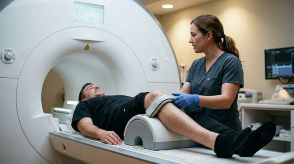
Positioning: The patient lies supine (flat on their back) on the motorized scanner bed. Coil Placement: A dedicated knee coil—a padded radiofrequency receiver shaped like a rigid boot or sleeve—is secured around the target joint to capture signals from the tissue. Entering the Bore: The table moves into the cylindrical magnet. For a knee scan, the patient enters feet-first, leaving the head and upper torso near or outside the edge of the scanner bore, which minimizes claustrophobic discomfort. Sequence Acquisition: The scanner generates loud clicking, knocking, and humming sounds as gradient coils rapidly switch on and off. Earplugs or noise-canceling headphones are provided. The patient must remain perfectly still during active acquisitions to prevent motion artifacts from blurring the images.
Post-Scan Processing and Image Delivery
Following sequence completion, the MRI technologist checks the image series for clarity before releasing the patient.
Digital Image Files: Patients can request a copy of their raw DICOM image files saved to a CD or accessible via a cloud portal immediately after the scan. Radiology Report: A board-certified radiologist reviews the slice series, documents structural findings, and drafts an official diagnostic report. The finalized report is sent to the patient via email or secure portal within 24 to 48 hours.